In an experiment investigating ways to engage optical Brain-Computer-Interfaces (BCIs) we used functional near infrared spectroscopy (fNIRS) and machine learning (support vector machines) to try to automatically classify ‘brain states’. (Given a time series pattern, what what the participant imagining doing: calculation, playing tennis, navigation, mental imagery, talking, singing?) Participants achieved between 55 and 90% accuracy across all tasks. (With the better trained participants achieving highest accuracies.) But this is with optimal conditions overall: awesome hardware, careful preparation, dark room etc…
From my experience to use fNIRS for our purposes seems like a great idea at first: The signal is similar to the hemodynamic signal that you get from big fMRIs, it can be better localized etc. But there are some important drawbacks that, in my opinion, make it impractical to use at home:
a) as FuChieh has already pointed out, it completely fails to deliver a meaningful signal on even the slightest of movements. As with all optical methods, this seems unlikely to be solved any time soon.
b) the signal to noise ratio (SNR) is very small: the changes in brain oxygenation you’re looking for are very small and you need optimal recording conditions to visible : little hair, (fair hair is better), dark room, no movement, optimal placement etc
c) the signal is very localized: you need to place your optodes directly over the ‘sweet spot’ in order to assess it correctly
d) optodes need to be pressed against the skin somewhat in order to give a good signal. this does get uncomfortable!
So basically, if you vary your measurement site by a cm, if you place the optode over a bit of hair, over a blood vessel, if you move, if there are adverse lighting conditions (or any bright light at all), your measurements will be corrupted.
However, IF you manage to have a clean signal, you WILL be able to see brain activation in the signal with your bare eye. For example if you sit still and put the optode just above your motor cortex and imagine to move the respective bodypart in a complex and controlled manner, you will see an increase in deoxygenated hemoglobin whenever you imagine movement, going back to baseline after termination of mental imagery within a couple of seconds. So I can definitely see it being used somehow, somewhere…
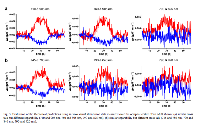
Just look at this beautiful signal here (raw data obtained during visual stimulation measured over the occipital cortex, Uludag et al, 2004):
 (see paper below)
A collegue of mine Kamil Uludag has looked at the problem of optimizing wavelengths for fNIRS:
http://spin.ecn.purdue.edu/fmri/PDFLibrary/UludagK_NI_2004_22_583_589.pdf
In essence he argues:
“The quality of the concentration changes’ assessment critically depends on
the wavelength combination used. Trying to optimize this combination, two
spectroscopic effects must be taken into account: cross talk and
separability. Cross talk between [oxy-Hb] and [deoxy-Hb] occurs
because the assumption made in the analysis—that there is a
homogeneous concentration change—does not hold true for the adult
human head. Separability—to be introduced in this paper—is a
measure for the degree of physical noise of the measurement that will
influence the noise of the concentration changes’ assessment. In other
words, high separability corresponds to a low noise with respect to the
concentration changes assessed”
“Cross talk leads to a ‘‘deformation’’, in some cases even an inversion of the
response direction, when the time courses of [oxy-Hb] and [deoxyHb] are
determined, while separability determines the noise level of the time course.”
“Our results (…) show that [oxy-Hb] and [deoxy-Hb] are determined more
accurately when using the wavelength combination 664 and 830
nm as opposed to 782 and 830 nm, and similarly that the
combination of 691 and 830 nm is superior to 780 and 830 nm.”
Check out the figure above: same task, same subject, same recording site, different wavelengths used.
No, sorry. Also, I’m really not sure about how accurate one can get with a DIY version. The originals use complicated lock-in amplifiers in order to achieve the SNR necessary for this. However, it might be easier if you’re looking at muscle instead of brain tissue?!
And yes it would be hard to get those readings, if not impossible.
That being said, I’d love to (help) develop a NIRS device for the enthusiasts. It’s definitely one measurement modality that’s still missing from our arsenal.
